
Ferris
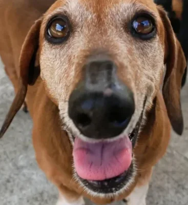
Ferris presented with advanced periodontal disease, bilateral oronasal fistulae, severe gum recession around both upper canine teeth, heavy plaque deposits and paradental ulceration.

Root canal therapy is a specialised endodontic treatment for teeth that are no longer vital or alive. When a tooth is damaged, such as by a fracture, the delicate dental pulp inside can become infected or diseased. This procedure removes the damaged pulp and seals the tooth, helping to save it and prevent further issues.
The dental pulp is a network of nerves, blood vessels and soft tissues within the tooth. It extends from the root to the crown, providing sensation, supporting dentine production and maintaining the tooth’s internal strength. As the tooth matures, the root canal gradually narrows. Root canal therapy maintains a non-vital tooth’s function as long as the gums, periodontal ligament and bone are healthy.
“Root canal therapy removes infected pulp and seals the tooth to help preserve its function.”

To restore the health of a tooth, we take the following steps:

We only recommend root canal therapy when a high likelihood of success exists, as not all teeth are suitable for this treatment. While we can’t guarantee outcomes due to pets’ unpredictable behaviour, the success rate is generally quite high.
Regular follow-up x-rays are crucial for assessing the progression and overall health of the treated tooth. While up to 90% of teeth treated with root canal therapy can last a pet’s lifetime, ongoing monitoring is essential for early detection of potential issues.
This is particularly important since pets cannot effectively communicate discomfort.
Root canal therapy is a specialised veterinary dental procedure used to treat teeth that are no longer alive due to infection or damage. It involves removing the diseased dental pulp and sealing the internal canal to prevent further infection. This allows the tooth to remain functional rather than requiring extraction.
The procedure begins with creating an entry point to access and remove infected tissue from inside the tooth. The canals are then carefully cleaned, shaped and dried before being filled with a sealing material to prevent reinfection. A final restoration is placed to protect the tooth, and in some cases, a crown may be recommended for additional support.
Root canal therapy for pets is considered when a tooth has been fractured or damaged in a way that affects the dental pulp. It is only recommended when there is a high likelihood of success, as not all teeth are suitable for treatment. The surrounding gums, bone and periodontal ligament must also be healthy to support the tooth.
While outcomes cannot be guaranteed due to factors such as a pet’s behaviour, the success rate is generally quite high. Up to 90% of treated teeth can last for the pet’s lifetime when properly managed. Careful case selection plays an important role in achieving these outcomes.
Pet root canal therapy is used to treat teeth that are no longer vital, removing infected pulp and sealing the tooth to help preserve its function.
The dental pulp supports tooth structure and sensation, and once damaged or infected, it must be removed to prevent further complications.
The procedure involves cleaning, shaping and sealing the root canal, followed by a restoration to protect the remaining tooth.
Follow-up x-rays are essential after treatment, as they help assess healing and monitor the long-term health of the tooth.
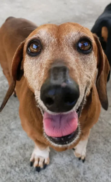
Ferris presented with advanced periodontal disease, bilateral oronasal fistulae, severe gum recession around both upper canine teeth, heavy plaque deposits and paradental ulceration.

Pip presented with a tight bite, a narrowed left maxillary diastema and linguoversion of the lower canine teeth.

Minnie underwent a thorough diagnostic process, including FIV/FeLV testing, biochemistry, and full-mouth radiographs.
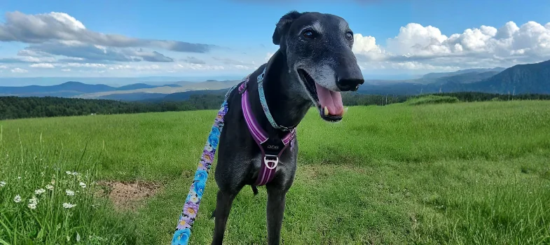
Ally is a beautiful Greyhound with complex cardiac risks who safely underwent anaesthesia for dental treatment, thanks to specialist anaesthetic management.
Ferris presented with advanced periodontal disease, bilateral oronasal fistulae, severe gum recession around both upper canine teeth, heavy plaque deposits and paradental ulceration.

Pip presented with a tight bite, a narrowed left maxillary diastema and linguoversion of the lower canine teeth.

Minnie underwent a thorough diagnostic process, including FIV/FeLV testing, biochemistry, and full-mouth radiographs.
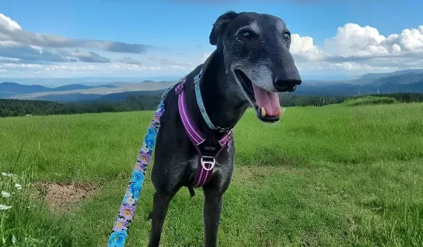
Ally is a beautiful Greyhound with complex cardiac risks who safely underwent anaesthesia for dental treatment, thanks to specialist anaesthetic management.


