
Ferris
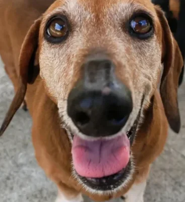
Ferris presented with advanced periodontal disease, bilateral oronasal fistulae, severe gum recession around both upper canine teeth, heavy plaque deposits and paradental ulceration.

Diagnostic imaging is a powerful tool that plays a pivotal role in oral care for animals. It provides veterinarians with a detailed view of structures that are hidden to the naked eye.
Dental X-rays are essential in modern veterinary dentistry. Intra-oral radiographs offer clear views of the tooth, pulp and surrounding structures, aiding in the diagnosis of issues that could otherwise go undetected.
We use digital radiography systems for easy image display, manipulation and enlargement on computer screens. This technology enhances diagnostic accuracy and safety by using lower doses of X-rays compared to traditional methods.
“Dental imaging reveals critical details that cannot be seen during a standard examination.”

Diagnostic imaging plays a key role in identifying and evaluating various dental conditions, including:
“Advanced imaging techniques provide deeper insight into complex dental and oral conditions.”

CT scans are necessary for a thorough examination of the anatomical structures surrounding the teeth. This advanced imaging technique provides detailed views of the head, including muscles, soft tissues, bones and joints.
Although CT scans take more time than standard dental X-rays, they offer crucial insights into complex conditions such as jaw fractures, Temporomandibular Joint (TMJ) disorders and oral cancers.
Having on-site CT scans improves our diagnostic capabilities and provides routine access to detailed imaging. This allows us to handle complex cases with greater precision and understanding as part of our regular procedures.
Dental imaging plays a central role in veterinary dental care by revealing structures hidden beneath the surface. It helps identify conditions that may not be visible during a physical examination, supporting a more complete understanding of animal oral health. This allows for more informed diagnosis and treatment planning.
Dental X-rays can detect a wide range of conditions, including bone loss from periodontal disease and missing teeth that are not visible during an exam. They are also used to assess root structures before extractions and identify issues such as tooth resorption in cats. This level of detail supports accurate evaluation of animal oral health.
Digital radiography allows images to be displayed, enlarged and adjusted on a computer screen for closer inspection. It uses lower doses of X-rays compared to traditional methods, which improves safety while maintaining image quality. This supports more precise diagnosis in veterinary dental settings.
CT scans are used when a more detailed view of the head and surrounding structures is needed. They are particularly helpful in assessing complex conditions such as jaw fractures, TMJ disorders and oral cancers. This type of imaging provides a deeper understanding of both hard and soft tissues.
Diagnostic imaging can help identify periodontal disease, hidden teeth, and structural issues within the roots. It is also used to investigate trauma such as jaw fractures and detect soft tissue abnormalities, including tumours. These insights are important for managing a wide range of animal oral health conditions.
Dental and oral imaging allows veterinary dental professionals to see structures that are not visible during a physical exam, supporting more accurate assessment of animal oral health.
Intra-oral radiographs provide detailed views of the tooth, pulp and surrounding areas, helping identify conditions that might otherwise go undetected.
Digital radiography systems enable easy image viewing and adjustment while using lower doses of X-rays, improving both safety and diagnostic accuracy.
CT imaging offers a comprehensive view of bones, joints and soft tissues, supporting the evaluation of more complex conditions affecting animal oral health.
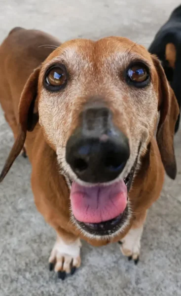
Ferris presented with advanced periodontal disease, bilateral oronasal fistulae, severe gum recession around both upper canine teeth, heavy plaque deposits and paradental ulceration.

Pip presented with a tight bite, a narrowed left maxillary diastema and linguoversion of the lower canine teeth.

Minnie underwent a thorough diagnostic process, including FIV/FeLV testing, biochemistry, and full-mouth radiographs.
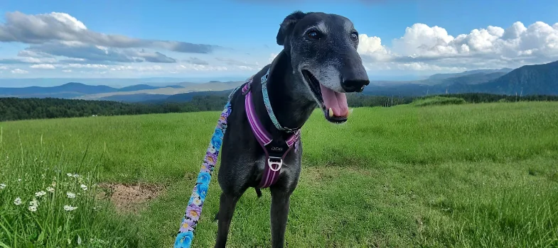
Ally is a beautiful Greyhound with complex cardiac risks who safely underwent anaesthesia for dental treatment, thanks to specialist anaesthetic management.
Ferris presented with advanced periodontal disease, bilateral oronasal fistulae, severe gum recession around both upper canine teeth, heavy plaque deposits and paradental ulceration.

Pip presented with a tight bite, a narrowed left maxillary diastema and linguoversion of the lower canine teeth.

Minnie underwent a thorough diagnostic process, including FIV/FeLV testing, biochemistry, and full-mouth radiographs.
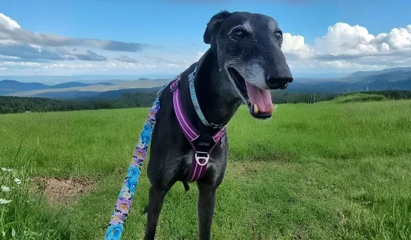
Ally is a beautiful Greyhound with complex cardiac risks who safely underwent anaesthesia for dental treatment, thanks to specialist anaesthetic management.


