Ferris Bilateral Oronasal Fistulae

Patient Information

Name: Ferris
Age: 13 years, 5 months
Breed: Dachshund
Gender: Male, Neutered

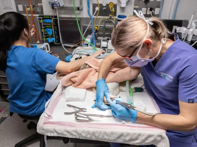
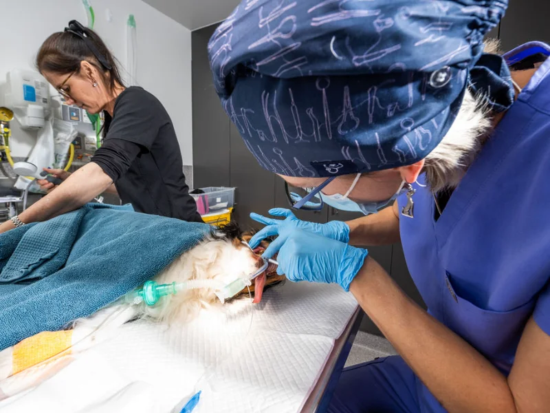
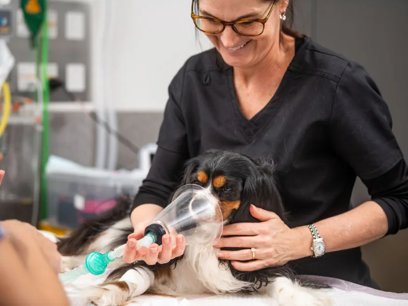
Ferris the Dachshund receiving specialist dental treatment at Advanced Animal Dentistry for severe periodontal disease and oronasal fistula repair
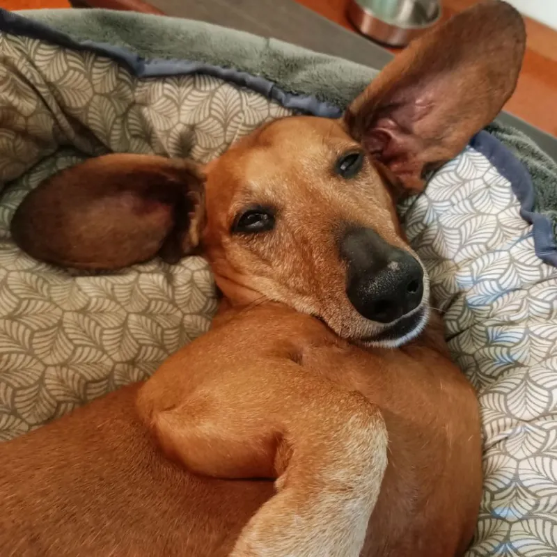
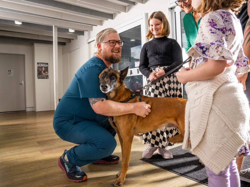
Ferris the Dachshund after successful treatment for severe periodontal disease and oronasal fistula at Advanced Animal Dentistry
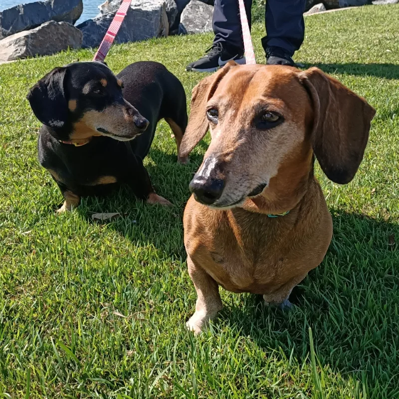
Ferris and his brother Bueller enjoying time outdoors after Ferris' successful dental treatment

"Due to the complications, our team recommended further pre-anaesthesia testing for Ferris rather than moving straight to anaesthesia."

Presenting Problem

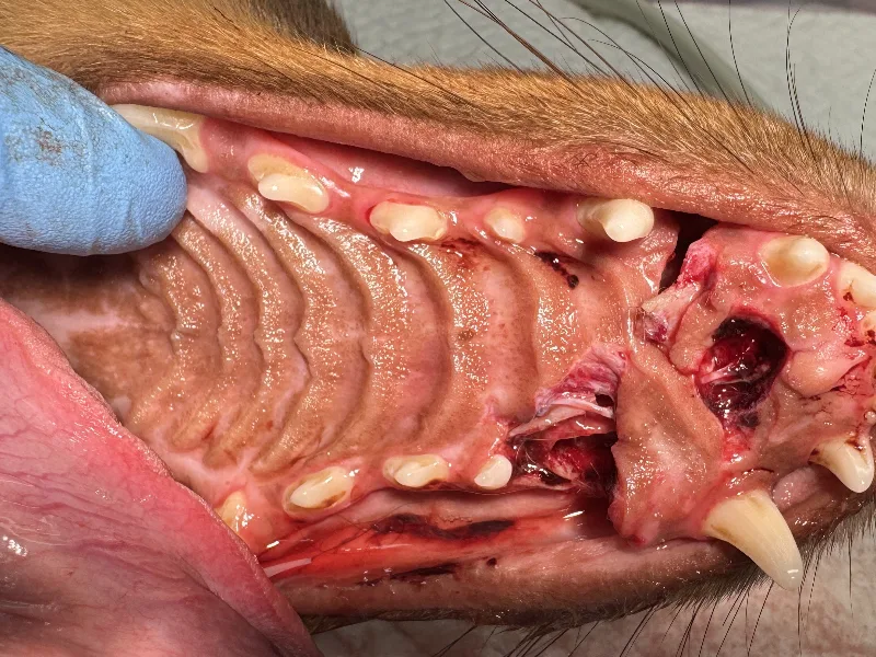
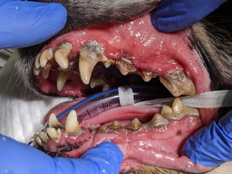
Ferris was referred by his regular veterinarian after his owners noticed worsening bad breath (halitosis), changes in the way he chewed because he was avoiding certain teeth and swelling near the right side of his muzzle. Initially, we found severe gum recession around both upper canine teeth, heavy plaque deposits and paradental ulceration. We were very suspicious of bilateral oronasal fistulae, which were later confirmed.

As Ferris is a senior dog with a moderate heart murmur, he required a cardiac assessment before proceeding.

Diagnostics and Treatment

Due to the complications, our team recommended further pre-anaesthesia testing for Ferris rather than moving straight to anaesthesia.

Pre-op medical management
We prescribed a two-week course of doxycycline to reduce inflammation and ulcerations around the fistula sites to give the surrounding tissue the best possible chance of healing successfully following the surgery.

Cardiac work-up
Because of the risks associated with Ferris’ age, his heart murmur and the fact he was facing a longer than normal procedure, he was referred for a pre-operative cardiology assessment. This confirmed mild (Stage B1) myxomatous mitral valve degeneration, which meant we could move forward with a highly tailored anaesthetic protocol.

Full oral assessment under general anaesthesia
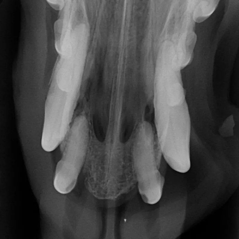
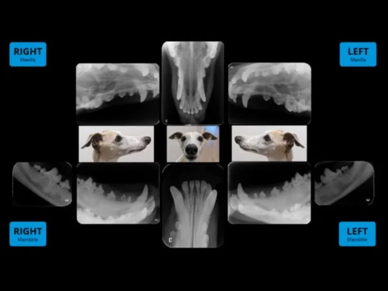
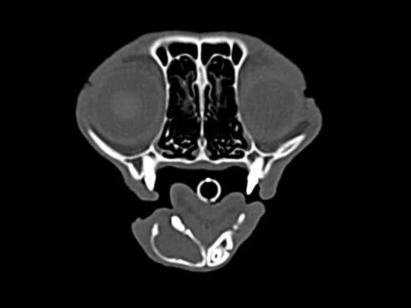
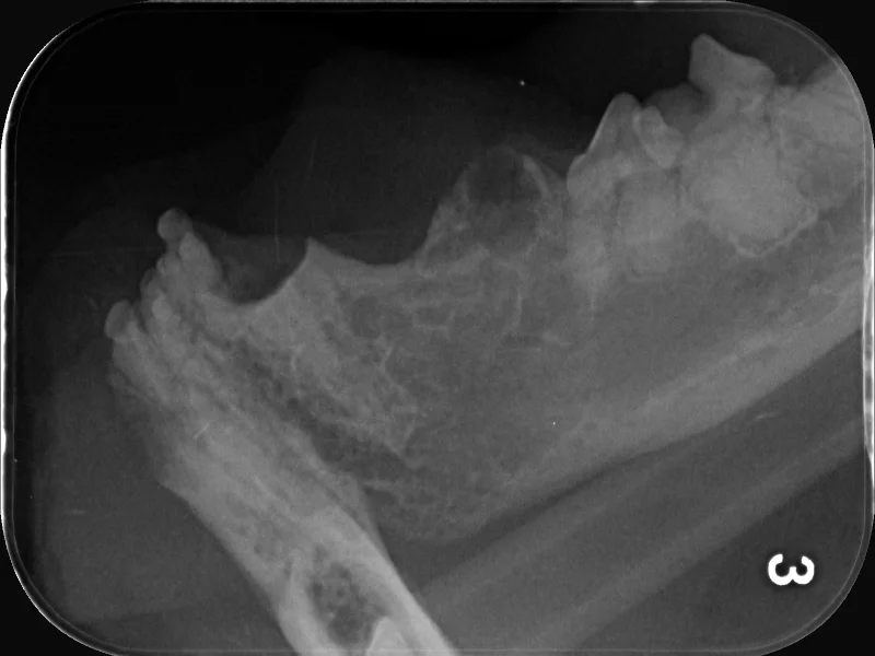
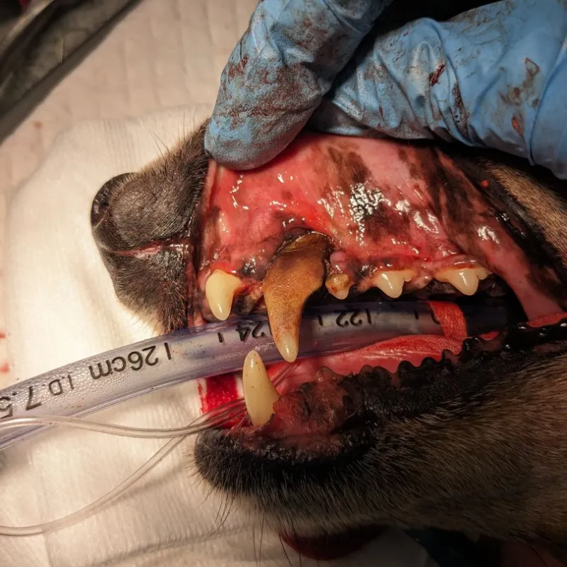
While under anaesthesia, we took dental radiographs that revealed extensive periodontal disease, including deep furcation involvement and several mobile teeth. We also confirmed bilateral oronasal fistula.

Extractions
We extracted 12 teeth, including both upper canines.

Bilateral oronasal fistula repair
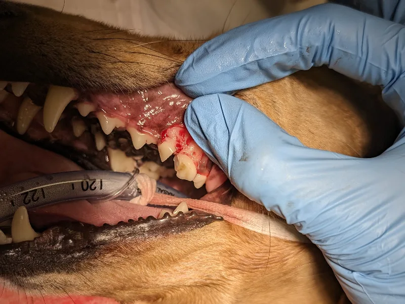
Next, we surgically repaired the upper canine oronasal fistula sites. Large mucoperiosteal flaps were raised, the nasal cavity flushed of debris and the surgical sites closed in a tension-free, layered closure. This was critical to preventing wound breakdown and recurrence of the oral-nasal communication.

We also used multimodal analgesia throughout the procedure, including regional nerve blocks. Ferris continued meloxicam and paracetamol at home during his recovery.

Dental radiograph showing advanced periodontal disease and bilateral oronasal fistulae
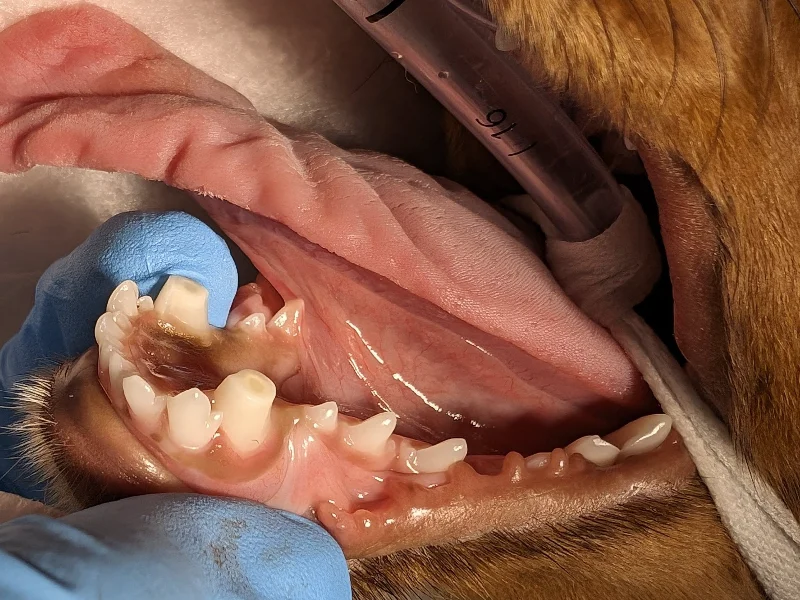
Dog's upper jaw after surgical removal of an upper canine tooth with sutures visible.
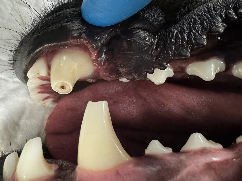
Close-up of Ferris' bilateral oronasal fistula caused by advanced periodontal disease before surgical repair

Outcome

Ferris made a smooth recovery. At his one-week recheck, both fistula repair sites were healing well with no evidence of breakdown and all sutures remained intact. By his two-week recheck, the sites had healed completely with no complications, and he was able to return to his normal diet and activity level. We also spoke with Ferris’ family about regular tooth brushing to help maintain his oral health.

"Careful surgical planning and meticulous technique were critical to repairing Ferris' bilateral oronasal fistulae."

Client Comment

“The surgery performed by Dr Tucker was life-changing for Ferris. His whole mouth was badly infected, he was unable to eat the foods that he enjoyed, and unfortunately, his dental condition was so poor that even his very experienced and capable vet was unable to treat him.

Since surgery, Ferris has thrived. He’s very active (as a 13-and-a-half-year-old Dachshund) and loves chewing on pig ears.
The post-surgery education about his oral care has really helped. Daily brushing or oral cleaning wipes, a change to the SPD diet and different chews have kept Ferris healthy. We’ve extended the same oral hygiene routine to his brother, Bueller, to help prevent future issues.

We are very grateful to Dr Tucker and Hayley for their excellent care of Ferris.”
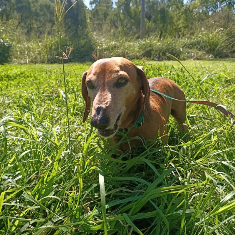
Brown dog wearing a green collar lying in tall green grass in a sunny field, looking at the camera.
Dental X-ray of the upper front teeth showing bone loss around the central incisors and root crowns with restorations present
Dental X-ray of the lower jaw showing tooth roots and surrounding bone structure on the left side of the image.
Dog's upper jaw after surgical removal of an upper canine tooth with sutures visible.
Relaxed brown dog with large ears lying on a patterned blanket in a cozy bed.

FAQs

Why was Ferris referred for a cardiac assessment before surgery?

Ferris was a senior dog, had not had recent pre-anaesthetic bloodwork and a heart murmur was detected during his examination. A pre-operative cardiology assessment confirmed mild (Stage B1) myxomatous mitral valve degeneration, allowing his anaesthetic protocol to be tailored to his specific needs.

What are the signs of an oronasal fistula in dogs?

Ferris’ owners noticed worsening bad breath, swelling near his muzzle and changes in the way he chewed because he was avoiding certain teeth. In his case, these signs, together with severe gum recession and ulceration, strongly suggested bilateral oronasal fistulae.

Why were dental radiographs important in Ferris' veterinary dental treatment?

Dental radiographs revealed the full extent of Ferris’ periodontal disease, including deep furcation involvement and several mobile teeth. This information guided the treatment plan, including which teeth required extraction.

Why were antibiotics prescribed before Ferris' surgery?

Ferris received a two-week course of doxycycline before surgery to reduce inflammation and ulceration around the fistula sites. Giving the surrounding tissue time to improve increased the likelihood of successful healing following the surgical repair.

How was Ferris' recovery managed after surgery?

Ferris recovered smoothly, with both fistula repair sites healing well and no evidence of wound breakdown. He continued meloxicam and paracetamol during recovery, returned to his normal diet and activity after two weeks, and his family received guidance on regular tooth brushing to help maintain his animal oral health.

Key Takeaways

Canine recession needs investigation

Noticeable gum recession may indicate an underlying oronasal fistula.

Reduce inflammation before surgery

Healthier tissue gives surgical repairs the best chance of healing.

Heart disease requires careful planning

A tailored anaesthetic plan helps manage higher-risk patients.

Surgical technique matters

Careful closure helps reduce wound breakdown and recurrence.

Next steps

For Pet Owners

Are you concerned about your pet’s dental health? Call 1300 866 848 or fill out the contact form. We’ll coordinate with your primary vet to ensure the best outcome.

For Vets & Nurses

We’re here to help. If you’d like to refer a patient, please call us at 1300 866 848 , complete the referral form or email us with a brief case summary, relevant radiographs and lab results.

For advice and treatment guidance, email us with a brief case summary, relevant radiographs and lab results.


Refer a Patient Contact Us

Advanced Veterinary Dental Services

A comprehensive range of dental and oral surgery treatments.

Contact Us    Refer A Patient
Complex Anaesthesia

We assess each patient individually, considering their health history, age and any concurrent medical conditions, to tailor the anaesthetic approach to their specific needs.

Surgical Extractions

Removing complex teeth safely and with minimal impact.

Vital Pulpotomy

A vital pulpotomy is a time-sensitive procedure used to preserve the life of a tooth when the pulp becomes exposed due to trauma or dental treatment.

Jaw Fractures

Jaw fractures in pets require precise treatment to restore both structure and function, with a strong focus on proper bite alignment.

Periodontal Treatment

Periodontal treatment goes beyond routine dental care to address the deeper damage caused by advanced gum disease.

Oral Surgery

Pet oral surgery can help treat dental problems, injuries, and other conditions affecting your pet’s mouth. Learn about the available procedures and how they can improve your pet’s comfort and oral health.

Dental and Oral Imaging

Dental and oral imaging helps veterinarians identify dental problems that may not be visible during a routine examination. Learn how these imaging techniques support accurate diagnosis and effective treatment for your pet.

Root Canal Therapy

Root canal therapy can help save a damaged or infected tooth while relieving pain in pets. Learn how this dental procedure works and how it can help preserve your pet’s natural teeth and oral health.

Gingivoplasty

This procedure involves reshaping gum tissue to guide the eruption of canine teeth into a correct position, preventing traumatic occlusion and avoiding more invasive treatments.

Crown Height Reduction

Reducing the height of misaligned canine teeth and protecting the exposed pulp with a filling helps prevent chronic pain, preserve jaw structure and avoid extraction.

Inclined Plane – Crown Extensions

Orthodontic appliances like inclined planes or crown extensions can reposition misaligned canine teeth, preventing trauma and preserving tooth function without altering their shape.

Dog Braces (Orthodontics)

To address mesioverted  (‘lanced’ maxillary canine teeth) and manage linguoversion when the maxillary diastema (the space between the third incisor and canine teeth) is too narrow.

Restorations

Restorative dentistry repairs damaged teeth in pets, often using crowns, veneers and fillings. These procedures restore dental structure and function, ensuring long-term oral health.

Professional Dental Cleaning

Thorough cleaning and examination under anesthesia remove plaque and calculus, preventing periodontal disease and ensuring long-term oral health in pets.

Mandibulectomies and Maxillectomies

These surgeries involve partial or complete removal of the jaw to treat malignant oral tumours. Treatment options vary and may include surgery, chemotherapy or radiation.

Crown Amputation for Tooth Resorption

This procedure, often used for tooth resorption, involves removing the tooth crown while leaving the resorbing root remnant intact. It’s less invasive and used in cases where traditional extraction is no longer possible. Regular follow-up is recommended.