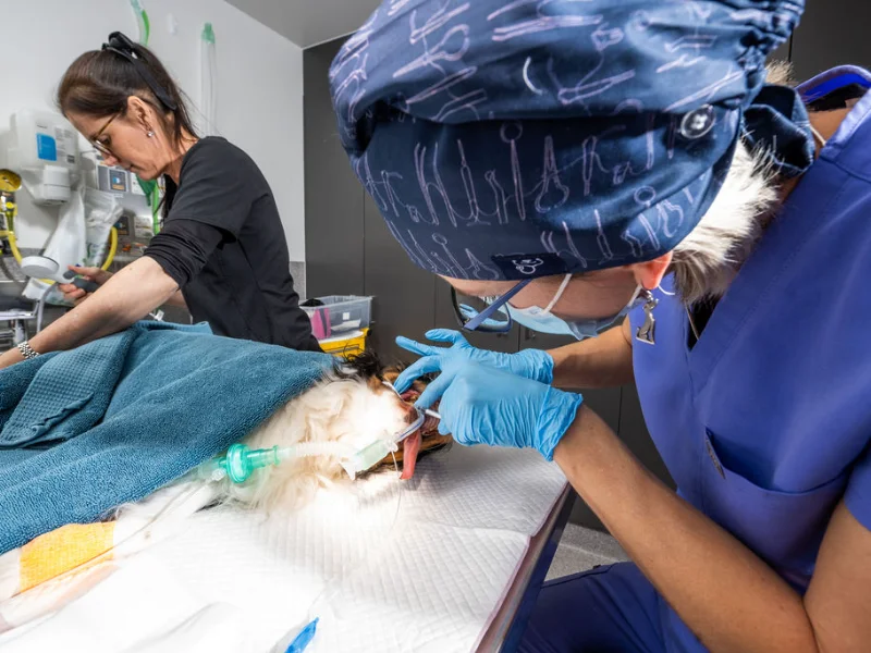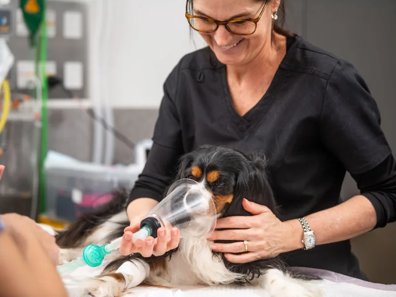
We assess each patient individually, considering their health history, age and any concurrent medical conditions, to tailor the anaesthetic approach to their specific needs.
Patient Information
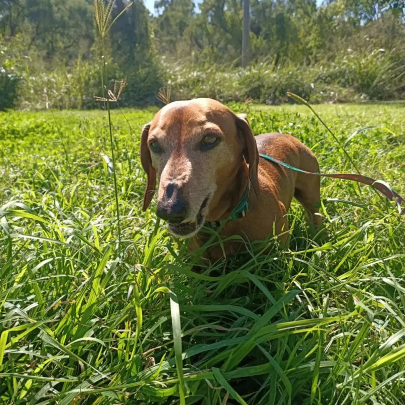
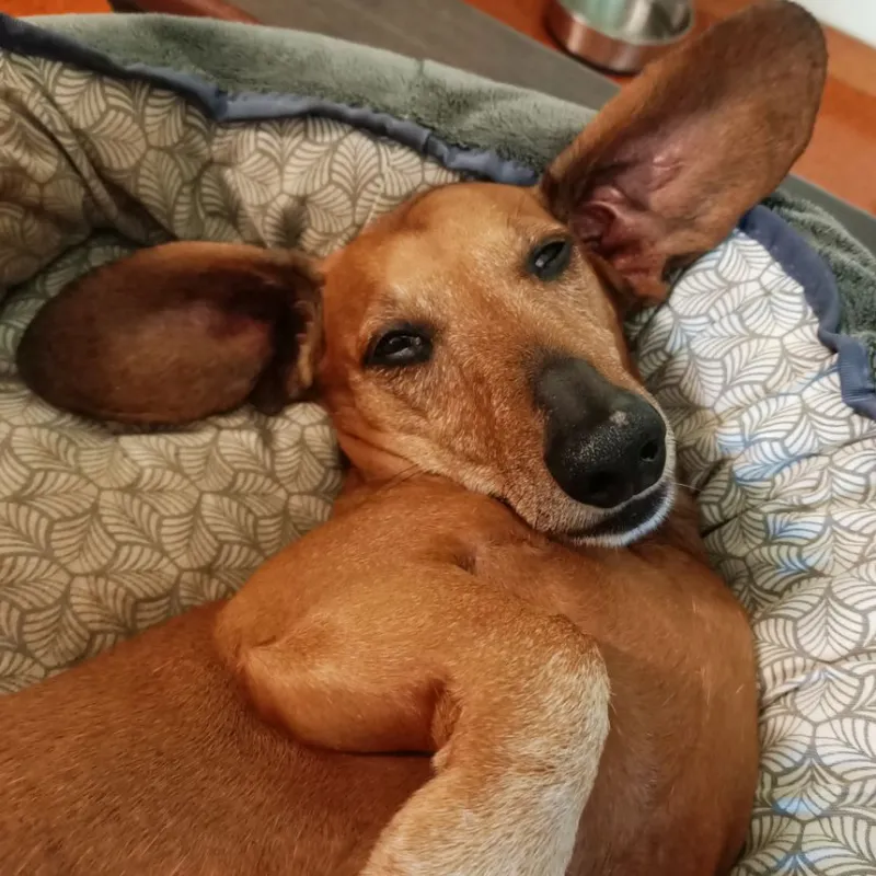
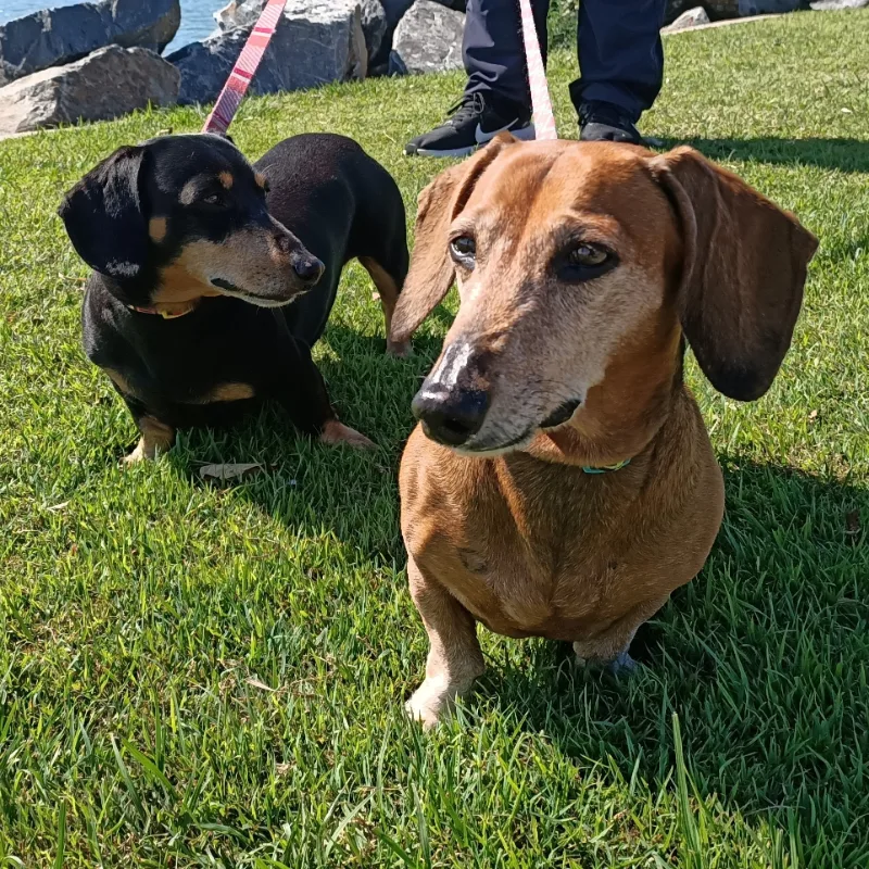
Name: Ferris
Age: 13 years, 5 months
Breed: Dachshund
Gender: Male, Neutered


"Due to the complications, our team recommended further pre-anaesthesia testing for Ferris rather than moving straight to anaesthesia."
Presenting Problem
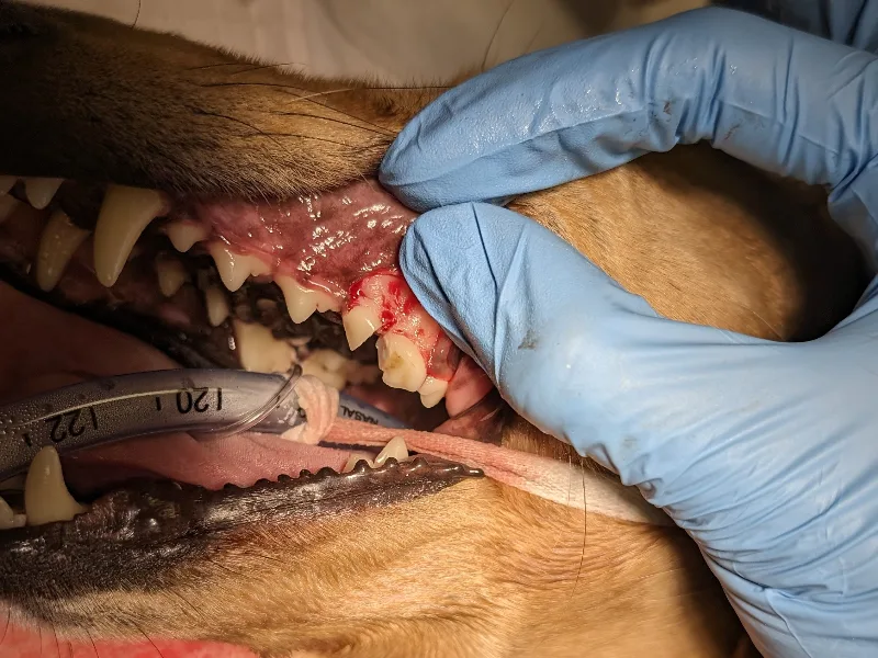
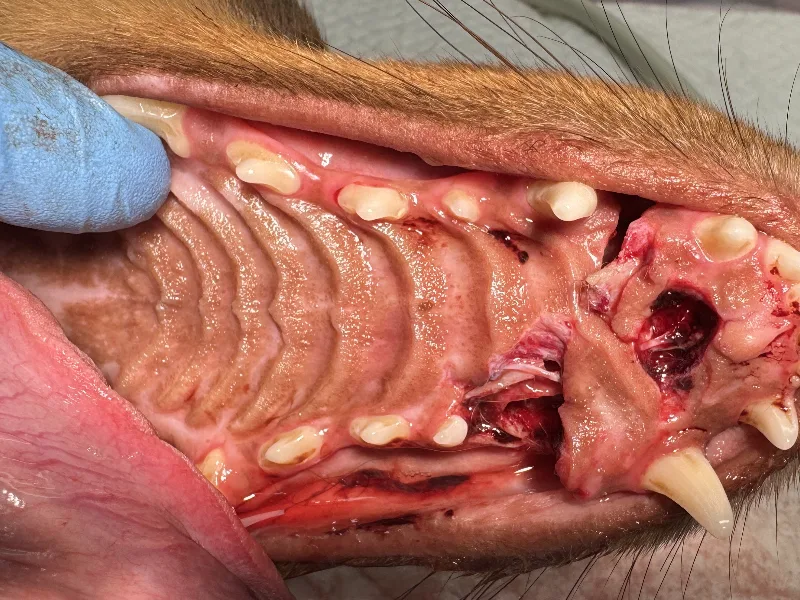
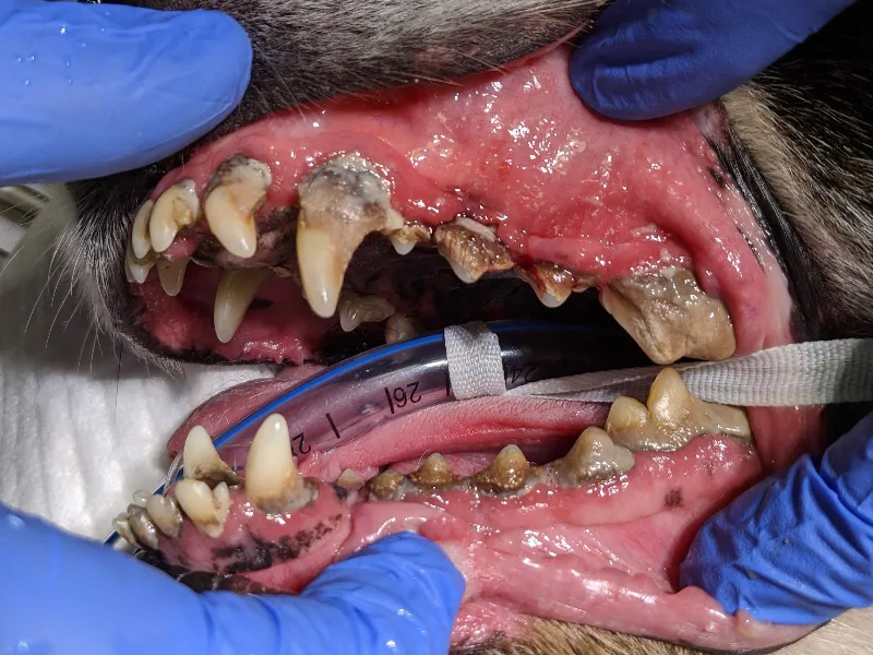
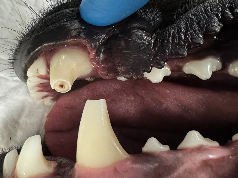
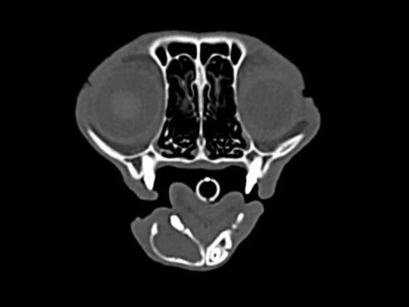
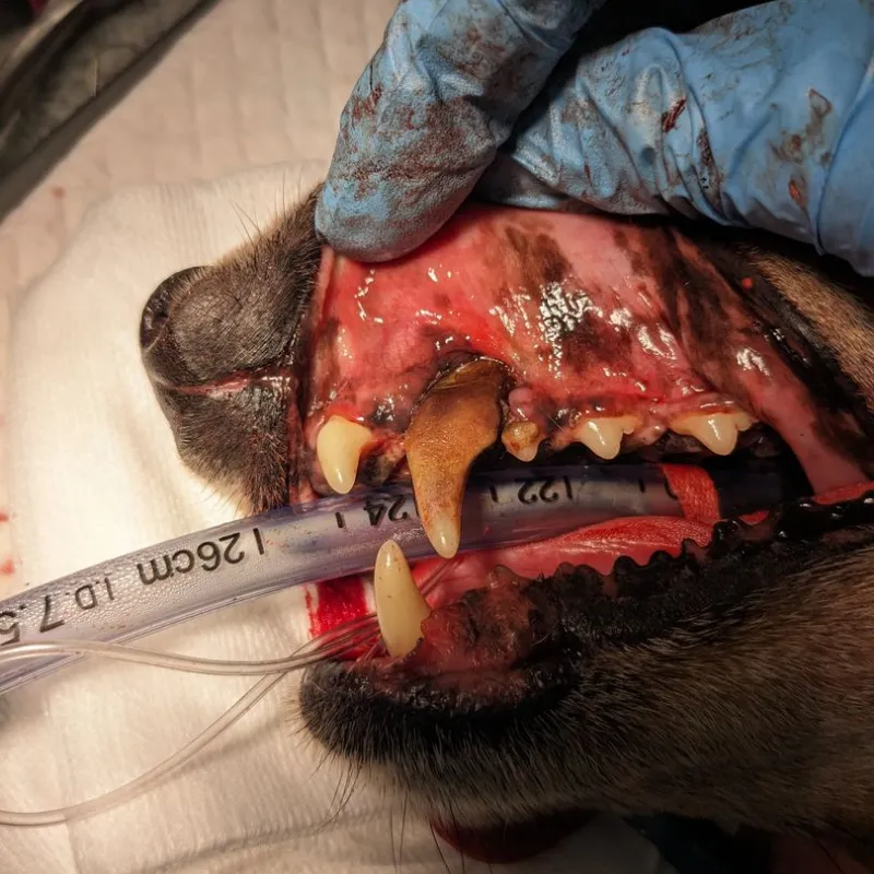
Ferris was referred by his regular veterinarian after his owners noticed worsening bad breath (halitosis), changes in the way he chewed because he was avoiding certain teeth and swelling near the right side of his muzzle. Initially, we found severe gum recession around both upper canine teeth, heavy plaque deposits and paradental ulceration. We were very suspicious of bilateral oronasal fistulae, which were later confirmed.
As Ferris is a senior dog with a moderate heart murmur, he required a cardiac assessment before proceeding.
Diagnostics and Treatment
Due to the complications, our team recommended further pre-anaesthesia testing for Ferris rather than moving straight to anaesthesia.
Pre-op medical management
We prescribed a two-week course of doxycycline to reduce inflammation and ulcerations around the fistula sites to give the surrounding tissue the best possible chance of healing successfully following the surgery.
Cardiac work-up
Because of the risks associated with Ferris’ age, his heart murmur and the fact he was facing a longer than normal procedure, he was referred for a pre-operative cardiology assessment. This confirmed mild (Stage B1) myxomatous mitral valve degeneration, which meant we could move forward with a highly tailored anaesthetic protocol.
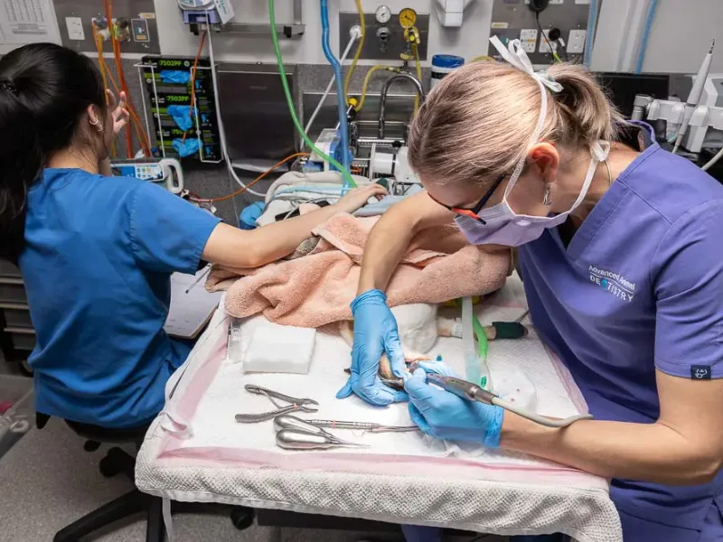
Full oral assessment under general anaesthesia
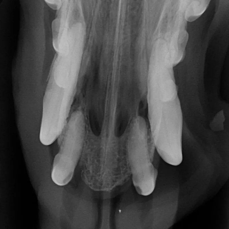
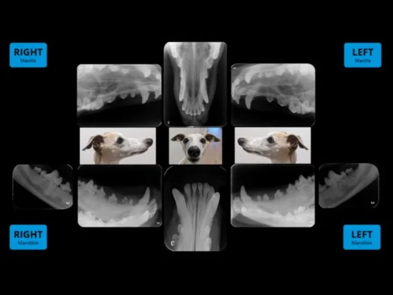
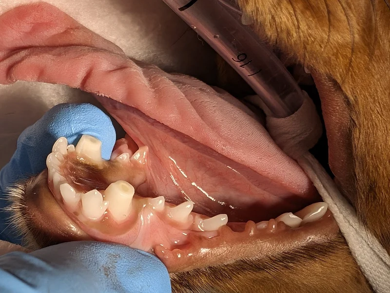
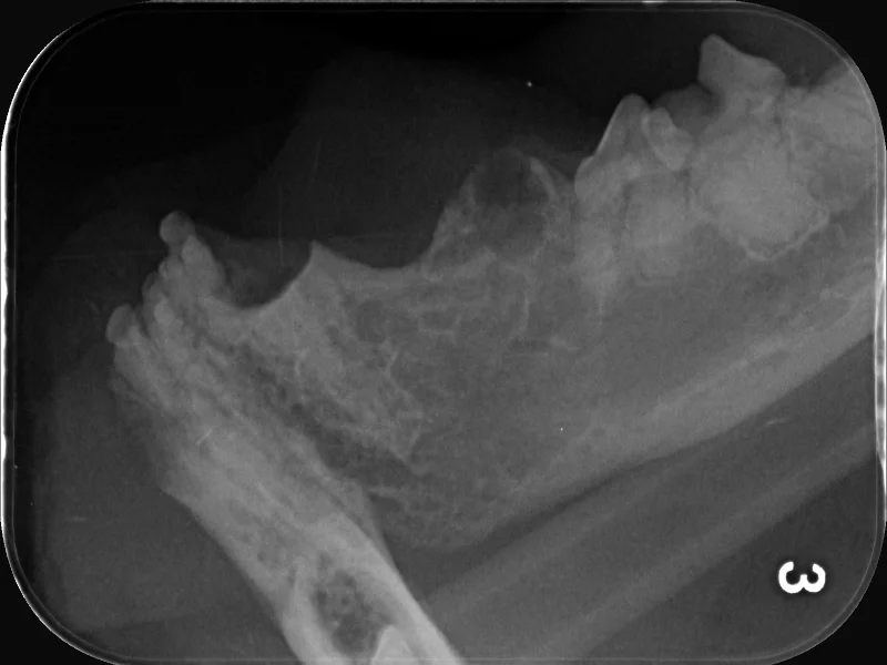
While under anaesthesia, we took dental radiographs that revealed extensive periodontal disease, including deep furcation involvement and several mobile teeth. We also confirmed bilateral oronasal fistula.
Extractions
We extracted 12 teeth, including both upper canines.
Bilateral oronasal fistula repair
Next, we surgically repaired the upper canine oronasal fistula sites. Large mucoperiosteal flaps were raised, the nasal cavity flushed of debris and the surgical sites closed in a tension-free, layered closure. This was critical to preventing wound breakdown and recurrence of the oral-nasal communication.
We also used multimodal analgesia throughout the procedure, including regional nerve blocks. Ferris continued meloxicam and paracetamol at home during his recovery.


Outcome
Ferris made a smooth recovery. At his one-week recheck, both fistula repair sites were healing well with no evidence of breakdown and all sutures remained intact. By his two-week recheck, the sites had healed completely with no complications, and he was able to return to his normal diet and activity level. We also spoke with Ferris’ family about regular tooth brushing to help maintain his oral health.
"Careful surgical planning and meticulous technique were critical to repairing Ferris' bilateral oronasal fistulae."
Client Comment
“The surgery performed by Dr Tucker was life-changing for Ferris. His whole mouth was badly infected, he was unable to eat the foods that he enjoyed, and unfortunately, his dental condition was so poor that even his very experienced and capable vet was unable to treat him.
Since surgery, Ferris has thrived. He’s very active (as a 13-and-a-half-year-old Dachshund) and loves chewing on pig ears.
The post-surgery education about his oral care has really helped. Daily brushing or oral cleaning wipes, a change to the SPD diet and different chews have kept Ferris healthy. We’ve extended the same oral hygiene routine to his brother, Bueller, to help prevent future issues.
We are very grateful to Dr Tucker and Hayley for their excellent care of Ferris.”